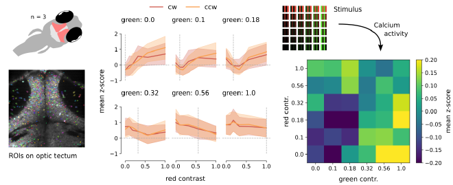
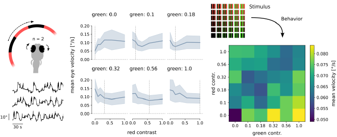
Experimental research has shown that zebrafish are less likely to perceive motion when the only moving element is color. Instead, it is the differences in brightness between moving stimuli that trigger a response (Orger and Baier, 2005). As part of a lab rotation project, we investigated how this behavior manifests in the optic tectum, the primary visual processing center in fish.
To achieve this, we employed two-photon calcium imaging to record the activity of direction selective neurons in the optic tectum of zebrafish larvae. Additionally, we simultaneously measured the optokinetic response, a behavioral indicator of motion perception.
The resulting dataset consisted of a z-stack of fluorescence microscopy images and the eye movements of the zebrafish embryo. After segmenting the cells with suite2p we analyzed the calcium activity (i.e., the luminance of each cell) and the eye velocities with respect to the stimulus that was shown to the fish.
The ensuing graphs illustrate a comparable pattern between the calcium
activities (neural signal) and eye velocities (behavioral output) for the same
stimulation conditions.

Our analysis suggested that the direction selective neurons in the optic tectum are likely colorblind, as they primarily respond to variations in brightness rather than color distinctions. As a result of this experiment, we have produced not only a poster but also a more comprehensive report, both of which can be found in the GitHub repository provided below.
A lab rotation with the systems neuroscience lab, University of Tuebingen, where we tested whether color contrast contributed to encoding motion in direction selective cells of the zebrafish optic tectum.
Image by Zeiss